X-ray manual : U.S. Army . <*5 lit w Fig. 3. Position for first exposure.. Fig. 4. Position for second exposure.200 HEAD EXAMINATIONS 201 moved from the front plane of the
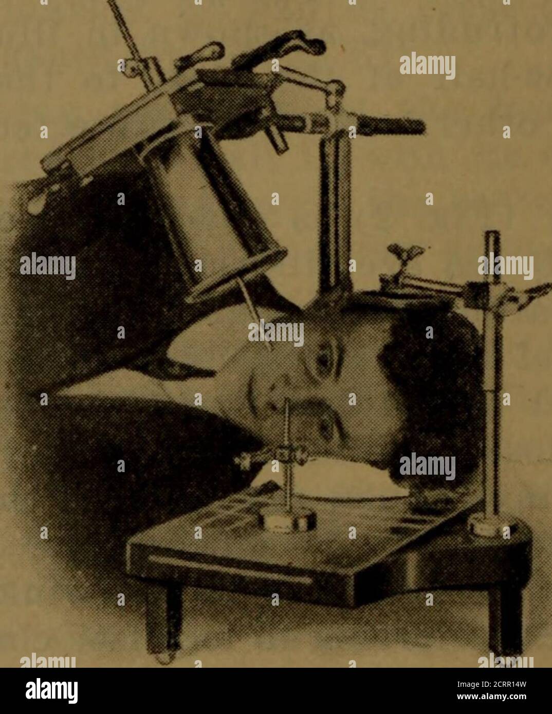
Download this stock image: . X-ray manual : U.S. Army . <*5 lit w Fig. 3. Position for first exposure.. Fig. 4. Position for second exposure.200 HEAD EXAMINATIONS 201 moved from the front plane of the cornea, and it shouldalso be borne in mind that the front of the cornea is 10millimeters in front of the shadow of the indicator-ball, asshown in your negatives. The tube is now centered overthe localizing ball and cone so that the shadows of thetwo will coincide (Fig. 3). Some object, such as a candle or a piece of whitepaper, that can be readily seen by the patient, should beplaced in alignment with the sights of the - 2CRR14W from Alamy's library of millions of high resolution stock photos, illustrations and vectors.

Guidelines for the Management of TDTs (3rd ed.)- English by Thalassaemia International Federation (TIF) - Issuu

May 2021 by Flyer & Aviation Publications - Issuu

Aviation visual Perception by batdelger - Issuu
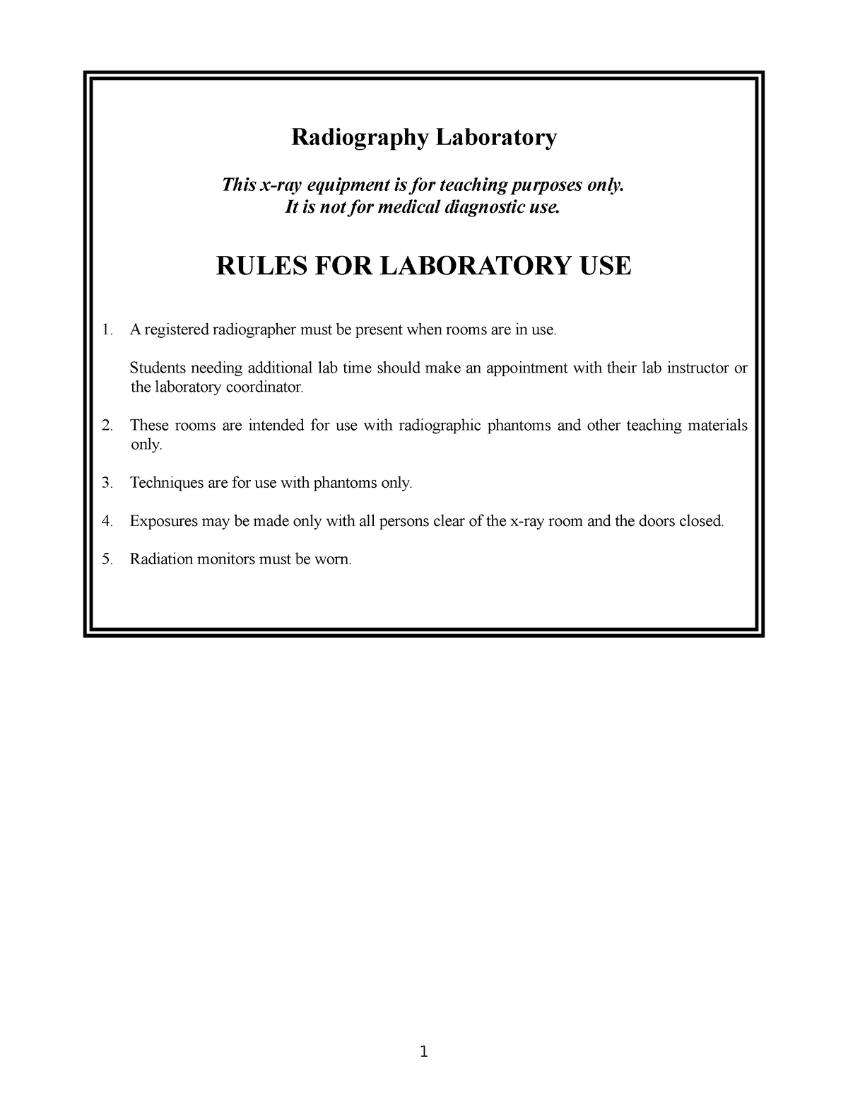
XRAY LAB Manual 2ND YEAR 2020 - Radiography Laboratory This x-ray equipment is for teaching purposes - Studocu
Solved Review the images in your lab manual to answer the

OAI X-ray Manual

Radiographic Exposure Technique

Abstract Book – 9th European Academy of Forensic Science Conference by NFC, Polismyndigheten - Issuu
Radiographic Exposure Technique

A comparative study of collimation in bedside chest radiography for preterm infants in two teaching hospitals - ScienceDirect

TJTES 2018-4 by KAREPUBLISHING - Issuu

lekarz wojskowy 2016 02 book kor en by Medycyna Praktyczna - Issuu

Ornl 2106 by The E Generation - Issuu

ICR 11.1 by Radcliffe Cardiology - Issuu

phywe-tess-phy-lep-en by SIDLAB,S. L. - Issuu